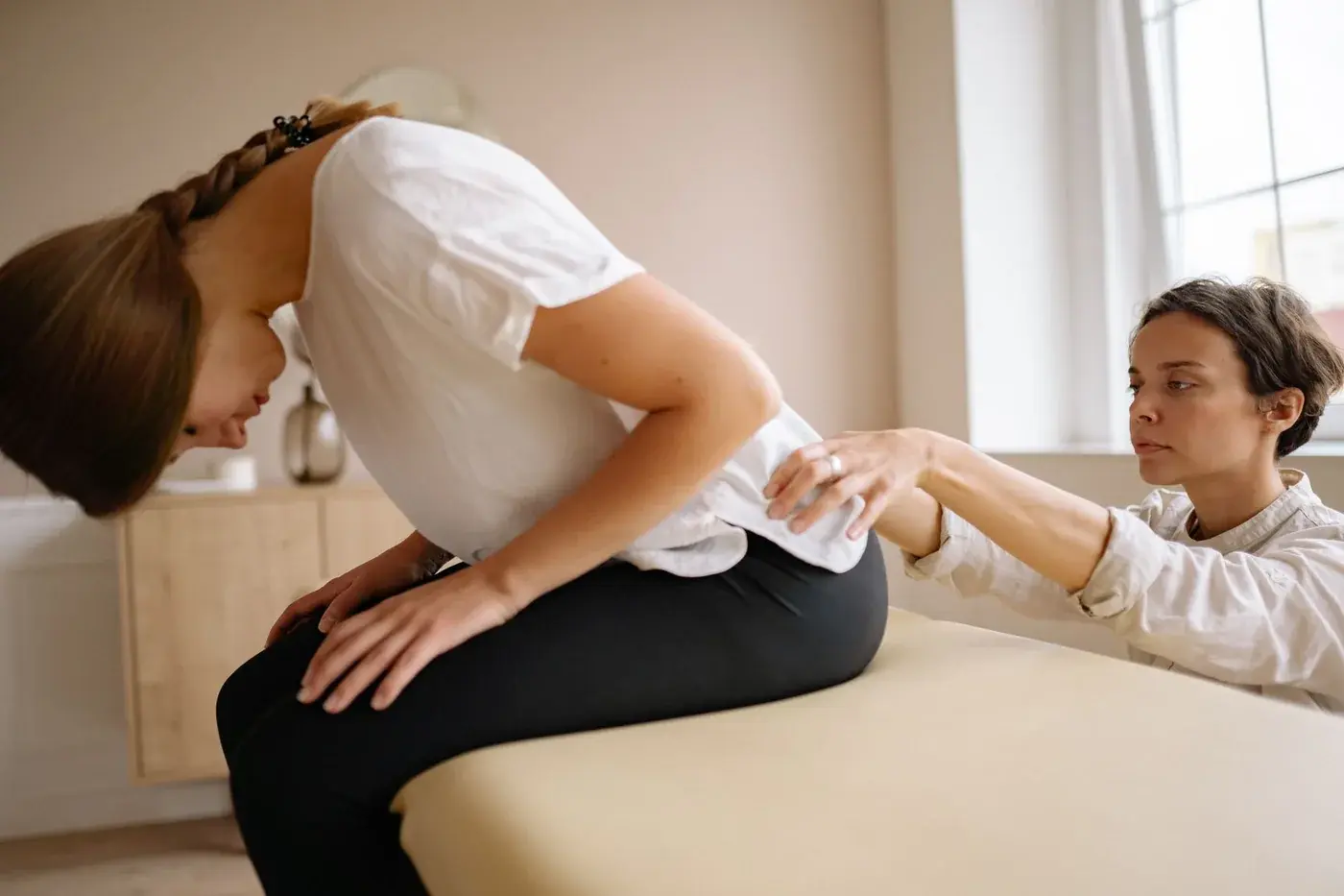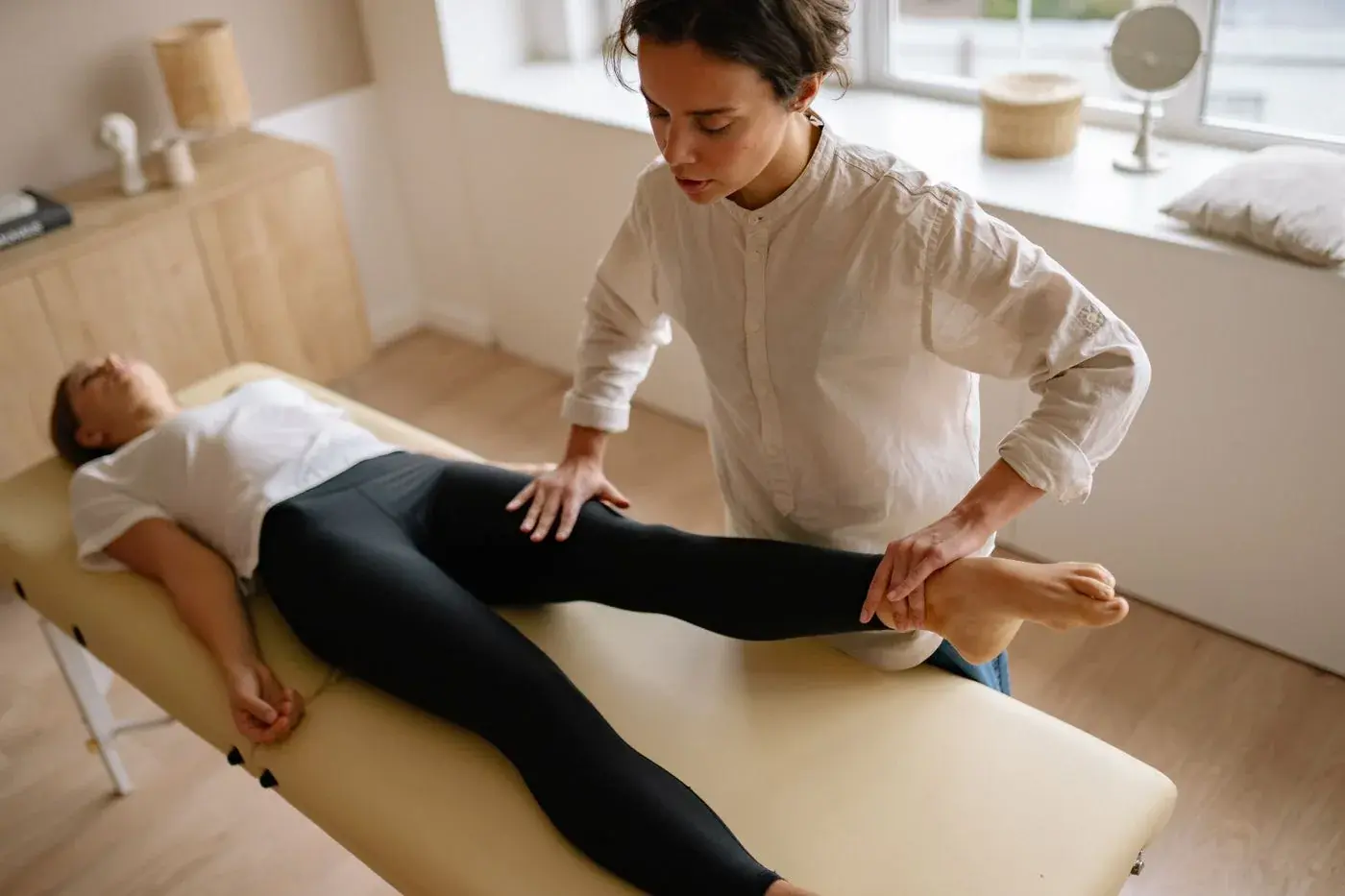
Hip pain and sciatica can overlap in confusing ways, especially when discomfort shows up in the groin, front of the thigh, side of the hip, or down the leg toward the knee or calf [4 (opens in a new tab), 9 (opens in a new tab)]. This guide outlines common symptom patterns that can suggest hip joint sources versus lumbar nerve–related pain, what day‑to‑day details to track before a pain‑clinic evaluation, and which warning signs require immediate emergency care rather than a routine appointment [3 (opens in a new tab), 6 (opens in a new tab), 7 (opens in a new tab)]. CMS Pain Management & Rehab in Sandy Springs evaluates back, hip, and leg pain and provides pain management and rehabilitation services [11].
Why hip pain and sciatica can feel similar
The hip joint and the lumbar spine share overlapping nerve pathways and movement demands, so pain from either region can present in the groin, buttock, thigh, or even the knee, making first‑pass sorting challenging without a careful history and exam [4 (opens in a new tab)]. Many adults have imaging findings in the hip or spine that do not cleanly match their symptoms, which is one reason clinicians rely on patterns over time, functional clues, and physical examination rather than imaging alone to localize the main pain generator [9 (opens in a new tab), 10 (opens in a new tab)]. When both the hip and the lumbar spine contribute (sometimes called hip–spine syndrome), coordinated evaluation helps set priorities for rehabilitation and activity planning [4 (opens in a new tab)].
Symptom patterns that can suggest hip joint pain
No single sign proves that the hip joint is the source, but certain clusters increase the likelihood and can be helpful to track for your visit [1 (opens in a new tab), 4 (opens in a new tab), 8 (opens in a new tab), 10 (opens in a new tab)].
- Location emphasis: Deep aching pain in the groin or front of the hip is a classic pattern for intra‑articular hip pathology and hip osteoarthritis, although it is not perfectly specific [1 (opens in a new tab), 4 (opens in a new tab), 10 (opens in a new tab)].
- Referred pain: Hip joint conditions can refer pain to the front or side of the thigh and sometimes to the knee, which can obscure the true source if knee imaging is normal [4 (opens in a new tab)].
- Stiffness and rotation limits: Loss of internal rotation and pain at the end range of hip motion are common in hip joint disease and may be noted by clinicians during an exam [1 (opens in a new tab), 4 (opens in a new tab)].
- Gait clues: A limp or a Trendelenburg‑type gait (pelvis dropping to the opposite side during single‑leg stance) can reflect hip abductor weakness or pain inhibition and may point clinicians toward hip involvement alongside other findings [4 (opens in a new tab)].
- Provocation maneuvers in clinic: Tests such as FADIR (hip flexion, adduction, internal rotation) and related intra‑articular provocation maneuvers may reproduce hip‑origin pain but have variable diagnostic accuracy across conditions and should be interpreted cautiously by clinicians in context [8 (opens in a new tab)].
Clues that can suggest lumbar nerve–related pain (sciatica)
Lumbar radicular pain (“sciatica”) typically reflects irritation or compression of a spinal nerve root, producing pain and sensory symptoms along a nerve distribution, but patterns can vary and require clinical correlation [7 (opens in a new tab), 9 (opens in a new tab)].
- Radiation pattern: Sharp, electric, or shooting pain running down the buttock and posterolateral thigh, often extending below the knee toward the calf or foot, is a common sciatica pattern, sometimes with numbness or tingling in a matching distribution [7 (opens in a new tab), 9 (opens in a new tab)].
- Cough/sneeze sensitivity: Worsening leg pain with coughing, sneezing, or straining can occur with radicular irritation due to transient increases in spinal canal or foraminal pressure [7 (opens in a new tab), 9 (opens in a new tab)].
- Exam maneuvers in clinic: A straight leg raise that recreates leg pain between roughly 30–70 degrees of elevation can support a radicular pain hypothesis; variants and crossed straight leg raise improve specificity when positive, but these are clinician‑performed tests and not suitable for self‑diagnosis [5 (opens in a new tab), 7 (opens in a new tab), 9 (opens in a new tab)].
- Neurologic changes: Dermatomal sensory loss, reduced reflexes, or focal weakness along a nerve root distribution can align with lumbar radiculopathy when found on exam, although not all patients show clear deficits [7 (opens in a new tab), 9 (opens in a new tab)].
Day‑to‑day function clues to jot down before your visit
Bringing a short, structured snapshot of your symptoms and function helps the clinic team localize the likely pain source and plan safer next steps. The items below are not tests and are not diagnostic by themselves; they organize observations that clinicians commonly weigh together with the physical exam [4 (opens in a new tab), 7 (opens in a new tab), 9 (opens in a new tab)].
- Pain map and timing: Note where pain starts, where it travels (groin, front/side/back of thigh, below the knee), what it feels like (aching, stabbing, burning), and whether nights or mornings are worse [4 (opens in a new tab), 7 (opens in a new tab), 9 (opens in a new tab)].
- Movement and position effects: Jot down positions or motions that predictably worsen or ease symptoms, including walking, pivoting, rising from a chair, stairs, prolonged sitting, or bending; also note whether coughing or sneezing spikes leg pain [7 (opens in a new tab), 9 (opens in a new tab)].
- Function and distance: Track how far you can comfortably walk, how long you can sit or stand, and whether one side feels weak or unsteady during daily tasks such as carrying groceries or climbing steps [4 (opens in a new tab), 9 (opens in a new tab)].
- Self‑care and sleep: Record whether tasks like dressing, getting into or out of a car, or lying on one side disturb your symptoms, and whether sleep is disrupted by the hip or leg [4 (opens in a new tab)].
- Neurologic sensations: Document any new numbness, tingling, or feeling of foot drag, and whether these sensations follow a strip of skin down the leg or sit mainly around the hip and groin [7 (opens in a new tab), 9 (opens in a new tab)].
- Recent illnesses, procedures, or risk factors: Note fevers, recent spinal or epidural procedures, immunosuppression, diabetes, or other infection risk factors, which can change the urgency of evaluation when combined with severe back pain or new neurologic findings [2 (opens in a new tab), 3 (opens in a new tab)].
Terms and exam maneuvers you may hear about in clinic (not for self‑testing)
Clinicians may mention named tests during evaluation. These maneuvers are interpreted in context and are not reliable when attempted at home [8 (opens in a new tab), 9 (opens in a new tab)].
- Straight leg raise: With you lying on your back, the examiner lifts a straightened leg; reproduction of typical radiating leg pain supports a radicular pain hypothesis when aligned with the history [5 (opens in a new tab), 7 (opens in a new tab), 9 (opens in a new tab)].
- Crossed straight leg raise: Raising the unaffected leg that reproduces pain down the symptomatic side is considered more specific but less sensitive for radiculopathy and is used alongside other findings [5 (opens in a new tab), 9 (opens in a new tab)].
- FADIR: Flexion, adduction, internal rotation of the hip can provoke intra‑articular hip pain; however, across pathologies the sensitivity and specificity vary widely, so the result must be weighed with the rest of the exam [8 (opens in a new tab)].
- Trendelenburg sign or gait: Pelvic drop on the opposite side during single‑leg stance or walking can indicate abductor weakness or pain inhibition often arising from hip conditions but can also appear with other causes; interpretation is clinical [4 (opens in a new tab)].
Emergency warning signs: seek immediate emergency evaluation, not a routine clinic visit
Certain symptoms point to conditions that require immediate emergency evaluation to protect nerve and overall health. If any of the following occur, emergency care is warranted rather than scheduling or waiting for a routine appointment [3 (opens in a new tab), 6 (opens in a new tab), 7 (opens in a new tab), 2 (opens in a new tab)].
- New trouble controlling bladder or bowels, new urinary retention, or new saddle‑area numbness — seek immediate emergency evaluation [3 (opens in a new tab), 6 (opens in a new tab), 7 (opens in a new tab)].
- Rapidly worsening leg weakness, especially if foot drop or knee buckling appears over hours to days — seek immediate emergency evaluation [3 (opens in a new tab), 6 (opens in a new tab), 7 (opens in a new tab)].
- Severe back pain with fever and any new neurologic deficits, or severe back pain after a recent spinal or epidural procedure, especially with diabetes, immune compromise, or intravenous drug use — seek immediate emergency evaluation due to concern for spinal epidural abscess [2 (opens in a new tab), 3 (opens in a new tab)].
- Back or leg symptoms with a history of known cancer plus new unexplained weight loss or night pain that wakes you from sleep — seek immediate emergency evaluation [3 (opens in a new tab), 6 (opens in a new tab)].
Limits, overlap, and why a clinic evaluation matters
Symptom location alone is imperfect: for example, some people with radiographic hip osteoarthritis do not report classic hip pain, and others with groin pain may have minimal radiographic change, so imaging and pain do not always align [10 (opens in a new tab)]. Similarly, lumbar MRI and nerve compression findings do not always match the severity or distribution of leg symptoms, and many adults have asymptomatic imaging changes, which is why diagnosis is primarily clinical [9 (opens in a new tab)]. Physical exam maneuvers for intra‑articular hip pathology show highly variable sensitivity and specificity across studies, and straight leg raise variants also have limits, so clinicians synthesize multiple clues rather than relying on a single test [8 (opens in a new tab), 5 (opens in a new tab), 9 (opens in a new tab)]. Hip and spine conditions can coexist and amplify one another, so a structured evaluation helps prioritize the main driver of pain and function limits for rehabilitation planning [4 (opens in a new tab)]. When red flags are present, imaging and emergency pathways are accelerated, whereas in the absence of red flags, many guidelines emphasize education‑first and time‑sensitive use of imaging for low back pain after clinician assessment [3 (opens in a new tab), 6 (opens in a new tab)].
Questions you can bring to your pain‑clinic visit
- Based on my pain map and function notes, which features lean more toward hip joint involvement and which suggest lumbar nerve irritation? How confident is that distinction today? [4 (opens in a new tab), 9 (opens in a new tab)].
- If the source is still uncertain, what examination steps or time‑based observations would help us narrow it down safely? [8 (opens in a new tab), 5 (opens in a new tab), 9 (opens in a new tab)].
- Do any of my symptoms qualify as red flags that change urgency or imaging decisions for low back or hip‑related pain? [3 (opens in a new tab), 6 (opens in a new tab)].
- If both hip and lumbar factors seem involved, how might we stage rehabilitation to address the most limiting driver of function first? [4 (opens in a new tab)].
- What signs should I monitor between visits that would prompt me to contact the clinic promptly versus seek immediate emergency evaluation? [3 (opens in a new tab), 6 (opens in a new tab), 7 (opens in a new tab), 2 (opens in a new tab)].
- If imaging is considered, how would the results change next steps, and what are the limits of imaging for correlating with symptoms in my situation? [9 (opens in a new tab), 10 (opens in a new tab)].
- Are there medication‑safety considerations I should understand as we discuss options, and how will any changes be coordinated with my prescribing clinician? (Do not make medication changes without the prescribing clinician.) [6 (opens in a new tab)].
FAQ
Can hip problems cause knee pain?
Groin and anterior thigh pain can originate from the hip joint and can also refer pain toward the knee, which can make knee pain feel primary even when the hip is the main source [4 (opens in a new tab)]. A clinic evaluation looks for patterns across location, motion, gait, and exam findings to clarify the driver [4 (opens in a new tab)].
If my thigh or leg pain patterns suggest sciatica but my MRI was called “normal,” could it still be a lumbar nerve issue?
Yes. Spine imaging findings and symptoms do not always match, and some people with clinical radicular pain have imaging that does not show a clear compressive lesion or shows changes that are common in people without symptoms [9 (opens in a new tab)]. Clinicians combine the history, exam, and time course to make the diagnosis; further testing is considered only when it would change management [3 (opens in a new tab), 9 (opens in a new tab)].
Is a home FADIR or straight leg raise a good way to tell hip pain from sciatica?
These maneuvers are best performed and interpreted by clinicians. FADIR and related hip provocation tests have highly variable accuracy across conditions, and straight leg raise variants have limits despite their usefulness when positive, so they are not recommended for self‑diagnosis [8 (opens in a new tab), 5 (opens in a new tab), 9 (opens in a new tab)]. Using your symptom and function notes is a practical way to prepare for a visit [4 (opens in a new tab)].
When is imaging needed right away for low back–related pain?
For low back pain without red flags, widely used appropriateness criteria advise against routine immediate imaging because early imaging rarely changes outcomes and can reveal incidental findings that do not match symptoms [3 (opens in a new tab), 6 (opens in a new tab)]. Red flags—such as new bladder or bowel dysfunction, saddle numbness, rapidly worsening weakness, or severe back pain with fever—shift the situation to immediate emergency evaluation [3 (opens in a new tab), 6 (opens in a new tab), 7 (opens in a new tab), 2 (opens in a new tab)].
Sources
- Does This Patient Have Hip Osteoarthritis? The Rational Clinical Examination Systematic Review - PMC (opens in a new tab)
- Spinal Epidural Abscess: A Review of Presentation, Management, and Medicolegal Implications - PMC (opens in a new tab)
- ACR Appropriateness Criteria® Low Back Pain: 2021 Update. (opens in a new tab)
- The Hip-Spine Connection: How to Differentiate Hip Conditions From Spine Pathology. (opens in a new tab)
- Extending the straight leg raise test for improved clinical evaluation of sciatica: validity and diagnostic performance with reference to the magnetic resonance imaging - PMC (opens in a new tab)
- Low Back Pain: Evaluation and Management - StatPearls - NCBI Bookshelf (opens in a new tab)
- Sciatica - StatPearls - NCBI Bookshelf (opens in a new tab)
- Sensitivity and Specificity for Physical Examination Tests in Diagnosing Prearthritic Intra-Articular Hip Pathology Are Highly Variable: A Systematic Review - PMC (opens in a new tab)
- Lumbar radicular pain - PMC (opens in a new tab)
- Association of hip pain with radiographic evidence of hip osteoarthritis: diagnostic test study - PMC (opens in a new tab)
- Pain Management in Sandy Springs, GA | CMS Pain & Rehab
When to Talk With a Clinician
For non-emergency questions, contact CMS Pain Management & Rehab to discuss your symptoms and care options with a qualified clinician. For emergency warning signs, call 911 or seek emergency care now rather than contacting the clinic.
Medical disclaimer: This article is for general education and is not a diagnosis or a substitute for individualized medical advice. Treatment options, risks, and eligibility vary; consult a qualified healthcare professional.
This article provides general information and is not an individual diagnosis or treatment recommendation. Ask your clinician how the information relates to your health. The accompanying stock photograph is illustrative.
Originally published Sep 30, 2026. Last updated Sep 30, 2026.