Why day-to-day patterns matter in lumbar spine pain (updated 2026-10-02)
Spinal stenosis vs herniated disc is a common comparison when leg pain, tingling, or weakness accompany low back symptoms. Both can narrow space around lumbar nerves, but they tend to affect daily function in different ways. Recognizing pattern clues—without self‑diagnosing—can help you prepare for a careful pain‑clinic evaluation and know which warning signs require immediate emergency care. Lumbar spinal stenosis often causes neurogenic claudication, a pattern of leg symptoms brought on by standing and walking that eases with sitting or leaning forward, while a lumbar disc herniation more often causes a focused radicular pattern (sciatica) that may worsen with coughing, sneezing, or prolonged sitting and can follow a single nerve’s territory in the leg [2 (opens in a new tab), 6 (opens in a new tab), 9 (opens in a new tab), 10 (opens in a new tab), 3 (opens in a new tab), 4 (opens in a new tab), 12 (opens in a new tab)].
Day-to-day pattern clues: lumbar spinal stenosis with neurogenic claudication
People with lumbar spinal stenosis commonly notice leg pain, heaviness, tingling, or numbness that starts or worsens with standing and walking and improves with sitting or leaning forward—the classic “grocery cart” sign. Symptoms can affect one or both legs and may feel more diffuse or patchy rather than tracing a single narrow line. Bending slightly forward, using a walker, or sitting can relieve symptoms because flexing the spine increases space in the spinal canal, while extension (standing tall, leaning back, downhill walking) may narrow it and provoke symptoms. Walking tolerance often decreases, and many people plan routes with places to rest. Back pain can be present, but leg symptoms typically dominate activity limits in neurogenic claudication [2 (opens in a new tab), 6 (opens in a new tab), 9 (opens in a new tab), 10 (opens in a new tab), 11 (opens in a new tab), 13 (opens in a new tab)].
Day-to-day pattern clues: lumbar disc herniation with sciatica symptoms
A lumbar disc herniation more often produces sharp or burning leg pain that can be worse than the back pain and may follow a single nerve root’s pathway (dermatomal pain), sometimes with numbness or tingling in the same territory. Sitting, bending, coughing, or sneezing can increase pressure in the disc and may aggravate symptoms. Some people recall a bend, lift, or twist before the onset, though not everyone has a clear trigger. On exam, clinicians often consider the straight leg raise test, which can reproduce radicular leg pain when the affected nerve root is irritated; interpretation depends on the full neurologic exam and history. Patterns of muscle weakness and reflex change, when present, can align with specific nerve roots. Many individuals with a lumbar disc herniation improve over time, though the course varies and requires individualized evaluation [3 (opens in a new tab), 4 (opens in a new tab), 12 (opens in a new tab), 8 (opens in a new tab), 14 (opens in a new tab)].
- Standing or walking makes leg symptoms worse, and sitting or leaning forward brings relief may be consistent with neurogenic claudication from lumbar spinal stenosis patterns; extension tends to aggravate, while flexion eases symptoms [2 (opens in a new tab), 6 (opens in a new tab), 9 (opens in a new tab), 10 (opens in a new tab), 11 (opens in a new tab)].
- Coughing, sneezing, or prolonged sitting worsens leg pain that tracks a single nerve territory may be consistent with a radicular pattern often associated with lumbar disc herniation; exam may reveal dermatomal sensory changes or a positive straight leg raise when clinically appropriate [3 (opens in a new tab), 4 (opens in a new tab), 12 (opens in a new tab), 8 (opens in a new tab), 14 (opens in a new tab)].
What to track before a pain-clinic visit
Thoughtful notes make a spine visit more productive and safer. Consider tracking the following observations for a short period, without drawing conclusions on your own. Bring the notes to your appointment so your clinician can interpret them in context [16].
- Walking and standing tolerance: note the number of minutes until leg symptoms begin; record whether sitting or leaning forward eases symptoms and how quickly [2 (opens in a new tab), 6 (opens in a new tab), 9 (opens in a new tab), 10 (opens in a new tab)].
- Positions and postures: list what clearly worsens or helps (for example, downhill walking worse vs level or uphill, leaning forward at a counter better) [2 (opens in a new tab), 6 (opens in a new tab), 9 (opens in a new tab), 10 (opens in a new tab)].
- Pain map and sensations: mark where pain, numbness, or tingling are felt; note if they trace a narrow line down the leg or feel broader or patchy. Include any dermatomal-feeling areas (for example, outer calf, top of foot) without diagnosing the nerve involved [3 (opens in a new tab), 4 (opens in a new tab), 12 (opens in a new tab)].
- Exertion or pressure triggers: note whether coughing, sneezing, or prolonged sitting predictably worsen leg pain [3 (opens in a new tab), 4 (opens in a new tab)].
- Functional impact: record changes in stair climbing, getting out of a chair or car, lifting groceries, or walking the dog [16].
- Perceived strength changes: jot down tasks that feel harder (such as going on tiptoes or on heels), without testing to fatigue. If you notice rapid change, see the emergency section below [3 (opens in a new tab), 4 (opens in a new tab)].
- Sleep and rest patterns: which positions allow you to settle symptoms; how long relief lasts after sitting or leaning forward [2 (opens in a new tab), 9 (opens in a new tab)].
- Medication and self-care history: bring a complete list of what you have already tried, any side effects, and allergies; do not make changes without the prescribing clinician [16].
- Prior records: collect earlier imaging reports, operative notes, and a current medication and conditions list; include the dates and locations where studies were done so staff can obtain images when needed [16].
What to bring to CMS Pain Management & Rehab for a spine-pain evaluation
Preparing a concise packet helps the clinical team focus on safety, function, and next steps. CMS Pain Management & Rehab in Sandy Springs emphasizes education-first, rehabilitation-forward care for spine pain. These items typically support an efficient visit [15, 16].
- A recent symptom and activity log summarizing walking/standing tolerance, posture effects, and recovery time after rest [2 (opens in a new tab), 9 (opens in a new tab), 16].
- A pain diagram showing both back and leg areas with numbness or tingling; note any dermatomal-feeling zones without drawing conclusions [3 (opens in a new tab), 4 (opens in a new tab), 12 (opens in a new tab), 16].
- A current medication and supplement list, allergies, and prior side effects; do not adjust medications without your prescribing clinician [16].
- Copies of recent imaging reports and dates; if available, bring digital image copies so your clinician can correlate findings with your exam [16].
- A brief timeline of prior treatments, physical therapy, procedures, or surgeries, and how they affected function [16].
- Work and daily activity notes: typical loads, positions, commute, and supports at home that influence pacing and mobility [16].
What clinicians may check during the exam (and why patterns matter)
A responsible spine evaluation blends history, neurologic exam, and, when indicated, imaging. For suspected lumbar disc herniation with sciatica symptoms, clinicians often consider the straight leg raise test, which can provoke radiating leg pain when the stretched nerve root is irritated; your clinician interprets it alongside dermatomal sensory changes, reflexes, and strength testing. For stenosis patterns, clinicians pay attention to posture-dependent changes, walking tolerance, and relief with flexion—the “grocery cart” sign—along with neuro exam findings. Exam responses help decide whether and how to correlate symptoms with a single nerve root or a multilevel narrowing pattern before discussing imaging or other steps [8 (opens in a new tab), 14 (opens in a new tab), 3 (opens in a new tab), 12 (opens in a new tab), 2 (opens in a new tab), 9 (opens in a new tab), 10 (opens in a new tab)].
Imaging and timing: how guidelines use red flags, duration, and exam to guide decisions
In lumbar spine pain, routine early imaging is generally discouraged when there are no red flags or severe or progressive neurologic deficits — seek immediate emergency care or call 911; many degenerative findings are common even in people without symptoms. When imaging is considered, it is usually because the history and exam point to a condition where results would change management or because red flags raise concern for conditions like infection, fracture, cancer, or cauda equina — seek immediate emergency care or call 911 syndrome. Decisions vary with individual risk factors and clinical judgment [1 (opens in a new tab), 5 (opens in a new tab)].
Lumbar and cervical imaging considerations differ; the points here address lumbar patterns and should not be applied to new neck symptoms. If you develop new or rapidly changing neck symptoms, a separate evaluation framework is used [1 (opens in a new tab), 5 (opens in a new tab)].
Emergency red flags: when to seek immediate emergency care
- New trouble passing urine, new urinary retention, or new loss of bladder or bowel control — go to the emergency department now or call 911 [1 (opens in a new tab), 3 (opens in a new tab), 5 (opens in a new tab), 9 (opens in a new tab)].
- New numbness in the groin or saddle area — go to the emergency department now or call 911 [1 (opens in a new tab), 3 (opens in a new tab), 9 (opens in a new tab)].
- Rapidly increasing leg weakness, a suddenly dropping foot, or inability to rise from a chair or walk that is clearly worsening over hours to days — go to the emergency department now or call 911 [1 (opens in a new tab), 3 (opens in a new tab), 5 (opens in a new tab), 9 (opens in a new tab)].
- Severe back pain with fever or feeling ill, especially with infection risks — go to the emergency department now or call 911 [1 (opens in a new tab), 5 (opens in a new tab), 9 (opens in a new tab)].
- A history of cancer with new severe back pain and neurologic changes — go to the emergency department now or call 911 [1 (opens in a new tab), 5 (opens in a new tab)].
- Significant trauma with new neurologic symptoms — go to the emergency department now or call 911 [1 (opens in a new tab), 5 (opens in a new tab)].
Limits and uncertainty: why patterns help but do not decide
Pattern clues are helpful, but they do not replace a focused clinical exam. Some people have both lumbar spinal stenosis and a disc herniation, or imaging that shows stenosis without fitting symptoms. Degenerative changes on MRI increase with age and do not always match pain or functional limits. Conversely, a person can have classic neurogenic claudication or dermatomal sciatica symptoms with subtle or multi-level imaging findings that need careful correlation. Your clinician’s task is to match history, exam, and, when appropriate, imaging to your goals and safety needs [1 (opens in a new tab), 4 (opens in a new tab), 10 (opens in a new tab), 11 (opens in a new tab), 5 (opens in a new tab)].
Questions you can take to your clinician
- Based on my walking tolerance, posture relief, and pain map, which diagnoses are you considering, and what alternatives are still on the table? [2 (opens in a new tab), 9 (opens in a new tab), 10 (opens in a new tab), 3 (opens in a new tab), 4 (opens in a new tab)].
- Which exam findings (for example, dermatomal sensory change, reflex patterns, straight leg raise response, or posture‑dependent symptoms) point more toward lumbar spinal stenosis versus a disc herniation in my case? [8 (opens in a new tab), 14 (opens in a new tab), 2 (opens in a new tab), 9 (opens in a new tab)].
- If imaging is considered, which study and why? How would the result change management, and what are the trade‑offs of waiting versus proceeding now? [1 (opens in a new tab), 5 (opens in a new tab)].
- What rehabilitation and activity‑pacing strategies typically fit my pattern, and how will we monitor function and safety over time? Please coordinate any medication decisions with my prescribing clinicians [5 (opens in a new tab), 7 (opens in a new tab)].
FAQ
Can spinal stenosis and a herniated disc happen at the same time?
Yes. Age‑related changes can narrow the canal while a focal disc herniation affects a single nerve root, so mixed patterns occur. That is why posture‑dependent claudication features can coexist with dermatomal sciatica features, and a careful exam plus selective imaging is often needed to clarify priorities [4 (opens in a new tab), 10 (opens in a new tab), 11 (opens in a new tab)].
If my at‑home straight leg raise seems positive, does that mean I have a herniated disc?
Not necessarily. The straight leg raise can be sensitive for radicular pain from a disc herniation, but it is not specific on its own and requires professional technique and interpretation alongside neurologic findings and history. Self‑testing is unreliable and can be misleading [8 (opens in a new tab), 14 (opens in a new tab)].
If my leg pain eases when I lean forward, is it definitely spinal stenosis?
Leaning forward or sitting to ease leg symptoms is a classic clue for neurogenic claudication from lumbar spinal stenosis, but no single sign proves the diagnosis. Other conditions can mimic this pattern, and some people with stenosis do not report clear posture relief. A full evaluation is needed [2 (opens in a new tab), 6 (opens in a new tab), 9 (opens in a new tab)].
How long should I wait before getting imaging if a herniated disc is suspected?
Imaging decisions depend on red flags, severity and progression of neurologic findings, and whether results would change management. Guidelines generally discourage routine early imaging for lumbar pain without red flags. Discuss timing and modality with your clinician [1 (opens in a new tab), 5 (opens in a new tab)].
Sources
- ACR Appropriateness Criteria® Low Back Pain: 2021 Update. (opens in a new tab)
- Spinal Stenosis and Neurogenic Claudication - StatPearls - NCBI Bookshelf (opens in a new tab)
- Radicular Back Pain - StatPearls - NCBI Bookshelf (opens in a new tab)
- Lumbar Disc Herniation - PMC (opens in a new tab)
- VA/DoD Clinical Practice Guideline: Diagnosis and Treatment of Low Back Pain - PMC (opens in a new tab)
- A Review of Lumbar Spinal Stenosis with Intermittent Neurogenic Claudication: Disease and Diagnosis - PMC (opens in a new tab)
- Noninvasive Treatments for Low Back Pain: Current State of the Evidence (opens in a new tab)
- Physical examination for lumbar radiculopathy due to disc herniation in patients with low-back pain - PubMed (opens in a new tab)
- Lumbar Spinal Stenosis - StatPearls - NCBI Bookshelf (opens in a new tab)
- Lumbar Spinal Stenosis: Pathophysiology, Biomechanics, and Innovations in Diagnosis and Management - PMC (opens in a new tab)
- Degenerative Lumbar Spinal Stenosis - PMC (opens in a new tab)
- Lumbar disc herniation: Epidemiology, clinical and radiologic diagnosis WFNS spine committee recommendations - PMC (opens in a new tab)
- Natural Course and Diagnosis of Lumbar Spinal Stenosis: WFNS Spine Committee Recommendations - PMC (opens in a new tab)
- Neurological examination of the peripheral nervous system to diagnose lumbar spinal disc herniation with suspected radiculopathy: a systematic review and meta-analysis - PubMed (opens in a new tab)
- Pain Management in Sandy Springs, GA | CMS Pain & Rehab
- What to bring to a back pain appointment | CMS Pain & Rehab
When to Talk With a Clinician
For non-emergency questions, contact CMS Pain Management & Rehab to discuss your symptoms and care options with a qualified clinician. For emergency warning signs, call 911 or seek emergency care now rather than contacting the clinic.
Medical disclaimer: This article is for general education and is not a diagnosis or a substitute for individualized medical advice. Treatment options, risks, and eligibility vary; consult a qualified healthcare professional.
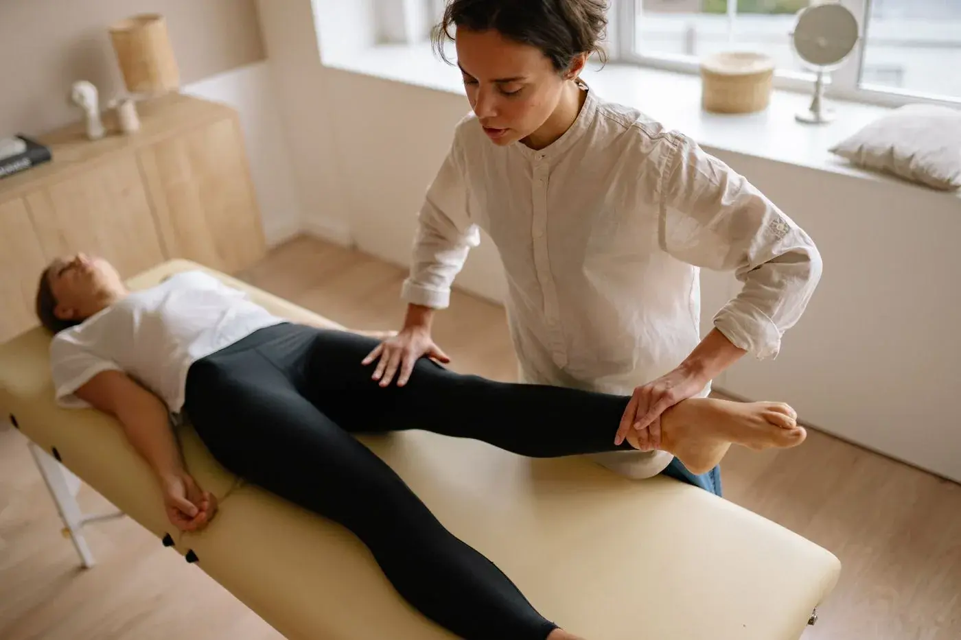
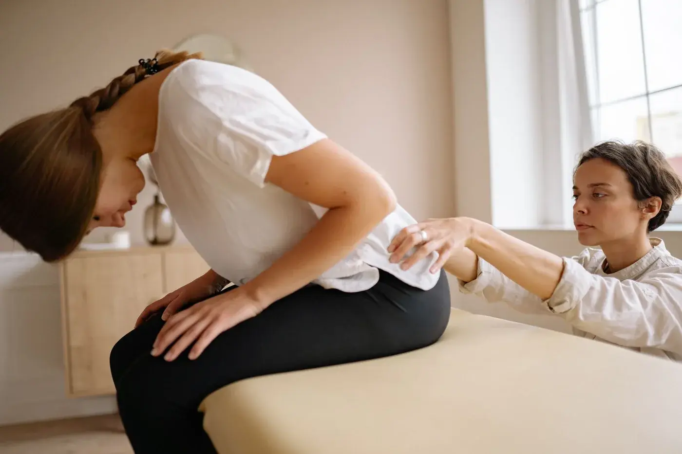
This article provides general information and is not an individual diagnosis or treatment recommendation. Ask your clinician how the information relates to your health. The accompanying stock photograph is illustrative.
Originally published Oct 2, 2026. Last updated Oct 2, 2026.